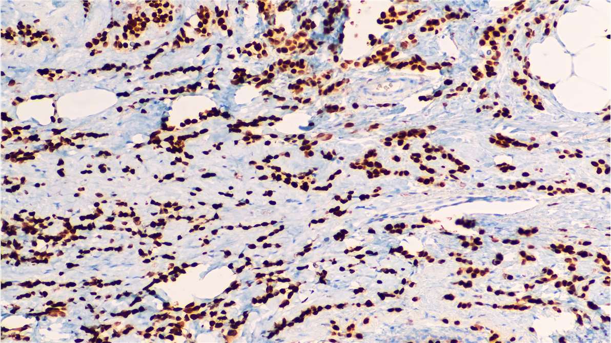
What is Immunohistochemistry?
Immunohistochemistry (IHC) is a microscope-based imaging technique that allows for the visualization of cellular components in a tissue sample. The unique feature that makes IHC stand out among many other laboratory tests is that it is performed without disrupting the histologic architecture, thus making it possible to assess the expression pattern of the molecule in the context of microenvironment.

How does Immunohistochemistry Work?
Tissue preparation
The tissue sample preparation is of crucial importance, because inadequate handling may disrupt tissue structure, resulting in reduced antibody binding affinity.
The most common processing for IHC is to prepare formalin-fixed paraffin-embedded (FFPE) tissue blocks. The purpose of formalin fixation is to produce chemical cross-linking of proteins within the tissue, which helps maintain tissue architecture. Another option is to cryopreserve (freeze) your samples, which may be a better choice if you are trying to detect a phosphorylated target.
Deparaffinization and rehydration
Where tissues are paraffin-embedded, it is essential that they are deparaffinized and rehydrated before being progressed through the IHC workflow. Deparaffinization is usually performed by immersing slides in xylene, whereas rehydration involves a series of incubations in decreasing concentrations of alcohol.
Antigen retrieval
Fixatives used to preserve the structural integrity of the tissue sample also have drawbacks in that they may bury the epitope your antibody is designed to recognize.
There are several ways to reveal epitopes that have been masked by fixation. These include proteolytic-induced antigen retrieval, which relies on an enzyme like proteinase K, or heat-induced epitope retrieval (HIER), which uses heat to break apart cross-linked bonds and unwind proteins.
Antigen retrieval is not required for frozen sections, but the sections should be air-dried for at least an hour prior to staining.
Protein blocking
Protein blocking step is required to reduce unwanted background staining. An ideal protein blocking agent is 5%–10% normal serum from the same species of secondary antibody. Other agents include protein buffers such as 0.1%–0.5% bovine serum albumin, gelatin, or nonfat dry milk.
Endogenous enzyme blocking
Blocking of endogenous peroxidase activity is indispensable when using the peroxidase-anti-peroxidase system for detection. 3% diluted hydrogen peroxide is widely used to block endogenous peroxidase activity. Likewise, endogenous alkaline phosphatase (AP), which is prevalent in frozen tissue, should be blocked with levamisole at a concentration of 10 mM.
Antibody application
There are two main types of antibodies, polyclonal antibodies which are heterogeneous population that bind to multiple different epitopes and monoclonal antibodies that all bind the same epitope. An advantage of using polyclonals for IHC is that they allow increased sensitivity through multi-epitope recognition, but they are available in only finite amounts and produce less consistent results due to batch-to-batch variability. In contrast, monoclonals can offer greater specificity and reproducibility, as well as being available in unlimited supply.
Direct or indirect detection is another important decision. During direct detection, the primary antibody is labeled, while indirect detection involves the use of a labeled secondary antibody. HRP- or AP- conjugated secondary antibodies are commonly used for IHC, where they are paired with dedicated HRP substrates and AP substrates for colorimetric detection. Among them, HRP-DAB is the most popular combination. The choice of HRP or AP mainly depends on antigen abundance and the level of endogenous enzyme present in the tissue section. Biotinylated secondary antibodies are also extremely popular for IHC and are routinely used in combination with enzyme-conjugated avidin or streptavidin.
Counterstaining
After the slide has been stained for the target of interest, it is often necessary to add a counterstain. Counterstaining labels all the cells and helps visualize IHC-labeled cells against the general morphology of the tissue. Counterstains used with chromogenic IHC include hematoxylin, nuclear fast red and eosin, while those used with fluorogenic IHC include 4',6-diamidino-2-phenylindole (DAPI), Hoechst33342 and propidium iodide.
What are the Applications of Immunohistochemistry?
IHC is applied across the whole spectrum of biological sciences. In biological research, IHC can visualize tissue organization, organ development, and apoptosis. IHC is also the preferred method for studying the expression of genetic material in the mitochondria of small tissue samples. In a medical setting, IHC can be used diagnostically to target specific markers of diseases and conditions. Finally, in drug development, IHC is used to verify the efficacy of a drug.