Histological staining plays a critical role in the visualization of tissue and cell structures. Living cells or fixed cells have very few inherent colors and only very small differences in the refractive indices of their component parts. By staining tissue sections, pathologists and researchers can view tissue morphology (structure) or look for the presence or prevalence of particular cell types, structures or even micro-organisms under a microscope.
Histological staining includes routine staining and special staining. Routine staining refers primarily to the hematoxylin and eosin (H&E) stain, while special staining is used to refer to alternative staining techniques outside of routine H&E stain to provide specific information researchers or pathologists need.
Routine Staining
Routine (H&E) staining is the cornerstone of tissue-based diagnosis. It stains the nucleus and a few other substances in blue, followed by counterstaining with an aqueous or alcoholic solution of eosin which colors cytoplasm and eosinophilic structures into various shades of red, pink, and orange for initial microscopic evaluation of the tissues. If applied properly, this technique can provide exceptional details of tissue structure and cell composition. This detail is necessary for tissue-based diagnosis, especially in the detection and classification of cancer. H&E staining is the main method in histology, and is considered to be the “gold standard” for brightfield microscopy.
Special Staining
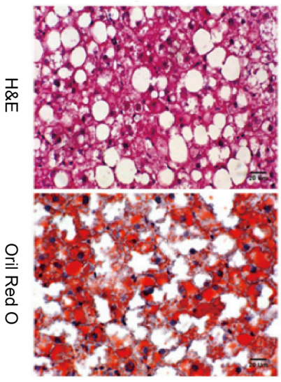
Based on routine staining methods, special staining techniques provide valuable information for the evaluation of numerous abnormal or disease conditions. They represent a variety of techniques and dyes with a more limited staining range, so they can be used to highlight certain features. In this way, special staining can be used for diagnosis or biomedical research, either to detect the presence of specific tissue structures or substances or to allow a more detailed morphological evaluation of these components and their distribution within a specimen. At Creative Bioarray, our Special staining includes but is not limited to:
|
|
|
|
|
|
|
|
|
|
|
|
|
|
|
|
|
|
|
|
|
|
|
|
|
|
If you do not see the stains you need on this list, please contact us and we will develop it for you. If you need any help in choosing a special stain for your project, please consult us before submitting a special stain request.