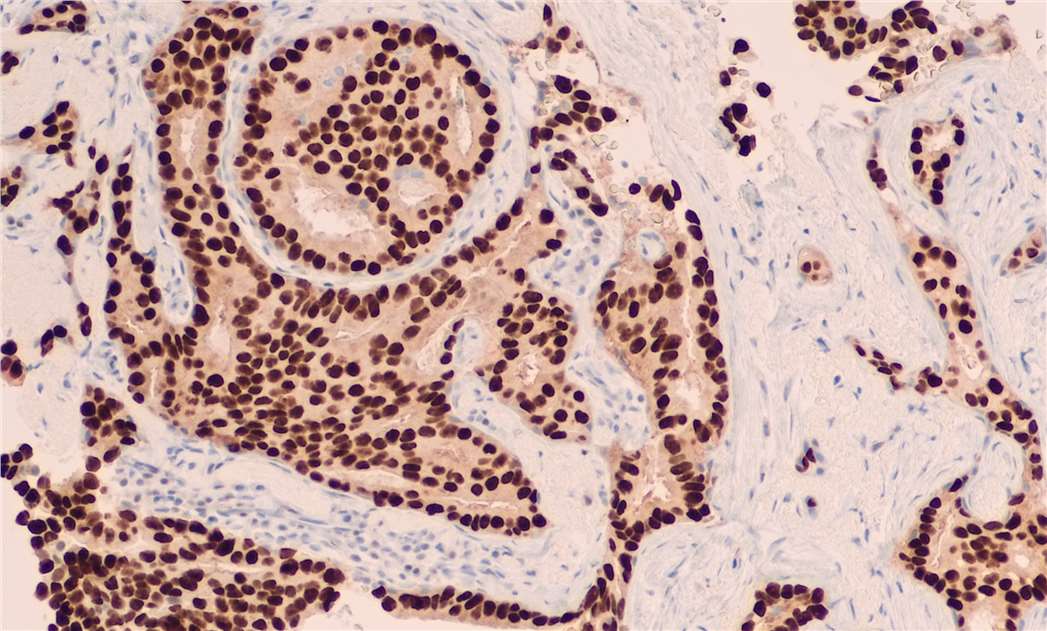
Immunohistochemistry (IHC) is a widely used biological technique that allows the end user not only to analyze the anatomy of the tissue of interest but also to visualize the expression, the localization and the intensity of a specific antigen on a tissue section. However, because IHC involves a greater number of protocol steps than many other immunoassay formats, there are more places where things can potentially go wrong. At Creative Bioarray, we are happy to provide simple but useful tips for improving daily tasks as well as the overall quality of your research.
How should I prepare my tissue samples?
Tissue samples for IHC staining can be either frozen or formalin-fixed paraffin-embedded (FFPE). Advantages of freezing include retention of antigen structure and faster processing times, while disadvantages include a loss of tissue morphology, and the need for cold storage. FFPE tissues are easier to handle, allow long-term storage at ambient temperature, and are generally more suitable for IHC, yet a drawback is that cross-linkage of proteins often require antigen retrieval. The method chosen will be influenced by factors including sample-handling capacity, available cold storage facilities, the need for sample archiving, and antibody compatibility with antigen retrieval.
Antigen retrieval – which induction system should I use?
FFPE samples typically require an antigen retrieval step prior to IHC staining, as the fixation process often result in antigen masking. Protease-induced epitope retrieval (PIER) involves treating samples with enzymes such as proteinase K, trypsin, or pepsin, but has a relatively low success rate and can damage tissue morphology. Heat-induced epitope retrieval (HIER), usually performed with microwaves or water baths, is more popular but is highly sensitive to timing, temperature, buffer, and pH. Antigen retrieval is less likely to be required for frozen tissue samples.
| PIER | HIER | |
| Temp | ~37℃ | ~95℃ |
| Conditions pH | Typically pH 7.4 | Citrate buffer of pH 6.0 or above. We recommend optimizing for best results. |
| Buffer | Neutral buffer solutions of enzymes such as pepsin, proteinase K or trypsin | Depends on pH required. Popular buffer solutions include Sodium citrate pH 6.0, EDTA pH 8.0 or Tris-EDTA pH 10.0 |
| Incubation time | 5-30 minutes | 10-20 minutes |
| Recommended antigens | Immunoglobulins, cytokeratins | No specific antigens |
| Precautions | Enzymatic retrieval can sometimes damage the morphology of the tissue. | Boiling can lead to tissue dissociation from the slide. Microwaving can result in uneven heat distribution. |
How to keep the background signal low? – blocking non-specific binding sites
Blocking nonspecific binding sites is key to preventing nonspecific staining. Blocking is typically done with normal serum from the species in which the secondary antibody was raised, although other blocking agents (such as BSA) are sometimes used. Increasing the concentration of blocking agent and incubation time might help to reduce nonspecific staining in your experiment.
Block endogenous peroxidases, phosphatases and biotin – what impact do these have?
Biotin presents endogenously in tissues and can have a profound impact when it comes to the visualization of these tissues. Avidin-Biotin blocking kit is important to prevent this. Peroxidases and phosphatase are also found endogenously. Use a suitable inhibition process to prevent their activity and the development of unwanted artefacts.
Are my antibodies and reagents suitable?
Sensitive and highly specific primary antibodies are essential to generate high-quality IHC data. Furthermore, one should never assume that an antibody that has been validated for frozen tissue will also be suitable for an FFPE sample, and vice versa.
Western blotting can often be used as the first line of specificity testing for a specific protein, as it can both quantify protein in your tissue, identify non-specific binding, and validate the specificity of a particular protein in combination with positive and negative controls. In many cases, polyclonal antibodies would be the most ideal to start a new IHC protocol because the variety of epitopes to which they bind allows for a stronger signal than that of monoclonal antibodies. But it's not only the antibodies, you will need to check you buffers, the antigen retrieval solutions and enzymes used. Carrying out a series of validation steps before starting an experiment is critical to prevent false positives or negatives occurring, and avoid waste on research funds.
Have I got the right detection system for IHC?
There are a range of systems available to detect the location and abundance of your protein using a fluorescent dye or chromogenic substrate. The greater the volume density of your target protein, the more suitable it will be for using a conjugated primary antibody to give a well-defined signal. However, if the antigen expression is not so high, using a secondary antibody can render a strong signal, because the signal may be amplified. For IHC, the most commonly used systems are streptavidin-biotin and avidin-biotin. Despite the best efforts to block endogenous biotin, non-specific staining can still occur with this system. There are nowadays alternatives for a crispier, more specific and more intense staining. These alternatives rely on enzyme-conjugated polymer reagents. They also have the added advantage of allowing you to reduce the amount of primary antibody being used.
Counterstaining – how does your counterstain contrast with your chosen chromogen?
Adding a counterstaining step after staining your slides with primary antibody provides better contrast to IHC data and facilitates insight into the exact location of target proteins within complex tissue structures. The choice of counterstain depends largely on whether you are using chromogenic or fluorogenic detection. While many counterstain options are available, the nuclear counterstains hematoxylin and DAPI are the most widely used for chromogenic and fluorogenic detection, respectively.
What are good IHC controls?
IHC, like all other assays, requires controls to confirm the accuracy and reliability of the staining pattern.
Antigen Controls - There should be both positive and negative antigen controls. The positive controls may be tissues known to express the protein under investigation. The negative controls are tissues known not to express the protein of interest. Using a negative control will help identify nonspecific bonding and false positives.
Reagent Controls - Reagent controls are used to ensure that staining is produced from the primary antibody staining the antigen and not from the detection system or the specimen. This can be determined using the detection system with diluent alone and no primary antibody.
For each experiment, it is also recommended to incubate tissue section with a non-specific isotype control antibody that matches the class and type of the primary antibody (if the primary antibody is a monoclonal antibody), but does not recognize the target epitope. Isotype controls help distinguish non-specific background fluorescence. If the primary antibody is a polyclonal antibody, incubate tissue section with a non-specific, species-matched polyclonal antibody.