H&E staining is a routine and essential staining technique used to study tissue samples in diagnostic, research and medical settings.
H&E staining has been used for at least a century and are still essential for recognizing morphologic changes in various tissue types. The staining has remained constant over the years, as it works well with a variety of fixatives and displays a broad range of cytoplasmic, nuclear, and extracellular matrix features.
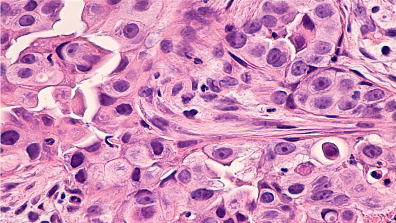
In a typical tissue, nuclei are stained blue, whereas the cytoplasm and extracellular matrix have varying degrees of pink staining. Well-fixed cells show considerable intranuclear detail.
The Principle of H&E Staining
The principle behind H&E staining is the chemical attraction between tissue and dye. Hematoxylin is a basic dye that imparts blue-purple contrast on basophilic structures, primarily those containing nucleic acid moieties such as chromatin, ribosomes and cytoplasmic regions rich in RNA. Acidic eosin counterstains the basic elements such as red blood cells (RBCs), cytoplasm, muscle and collagen in varying intensities of pink, orange and red.
Progressive, Modified Progressive and Regressive H&E Staining
There are typically three types of H&E staining: progressive, modified progressive and regressive.
Progressive H&E Staining
Progressive H&E staining is a gradual staining process where the histologist leaves the tissue in the staining solution long enough to achieve the desired endpoint. Progressive H&E staining allows for higher result intensity due to the longer staining process.
Mayer's and Gill's hematoxylins are naturally progressive, making them typical dyes for progressive H&E staining. Histologists often use them as nuclear counterstains for immunohistochemistry and special stains. Harris hematoxylin is another commonly used hematoxylin, especially for progressive staining of cytology specimens.
Regressive H&E Staining
Regressive H&E staining is a quicker staining process than progressive H&E staining. Histologists deliberately overstain the tissue until all of its components are dye-saturated. Then, they de-stain the tissue until it reaches the desired endpoint.
Histologists prefer regressive staining when very clear differentiation of tissue elements is required, as this process produces customized staining intensity. Harris hematoxylin is the commonly used hematoxylin for regressive staining, but Delafield's, Ehrlich's and Mayer's hematoxylins can also be used.
Modified Progressive H&E Staining
Rather than removing excess stain from the nuclei, modified progressive H&E staining uses a mild differentiator to remove background staining. What makes a stain progressive is the presence of an altered nuclear stain — not a differentiator. Similar to progressive staining, the differentiator should not be too strong. Otherwise, the nuclei will be stained too pale to read properly.