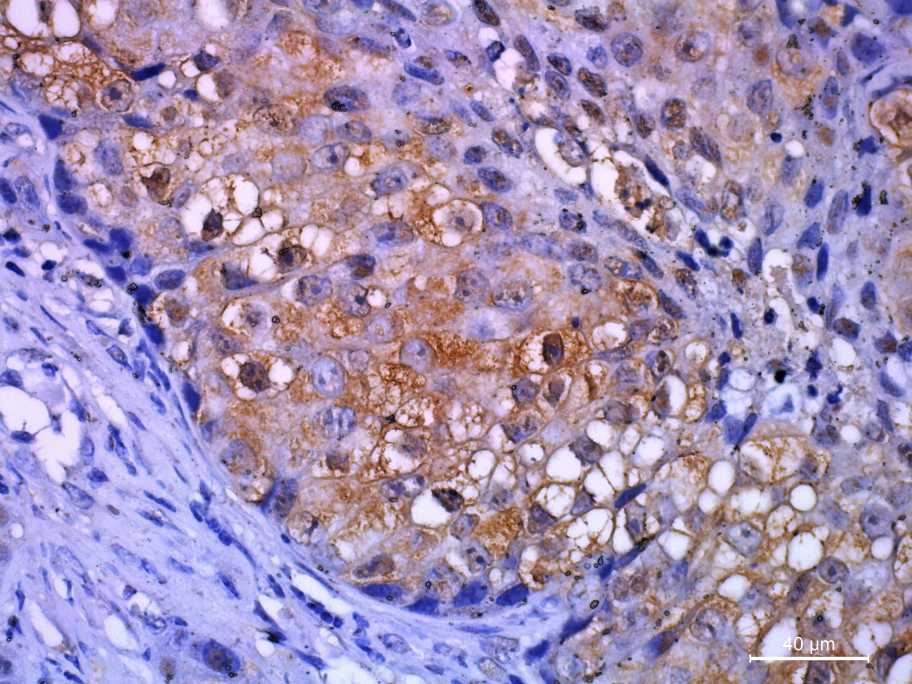
Immunohistochemistry (IHC) is a widely used technique that allows end user not only to analyze the anatomy of the tissue of interest, but also to visualize the expression, localization, and intensity of a specific antigen on a tissue section. While every immunoassay workflow requires researchers to make certain choices, IHC takes things that much further. In addition to deciding between direct or indirect detection, choosing whether to use enzyme-labeled or fluorophore-labeled antibodies, and determining if additional signal amplification is required, researchers performing IHC must also consider whether they will work with frozen or formalin-fixed paraffin-embedded (FFPE) tissue and if epitope retrieval is necessary for antibody reagents to access their targets. Each step of the IHC workflow contributes to the final result.

Frozen or paraffin sections?
The most common histological preparation technique is formalin-fixed, paraffin-embedded tissue. Some epitopes are more sensitive to fixation and embedding than others and can be masked upon addition of affinity reagents. Various antigen retrieval methods exist to unmask a given epitope. These are not applicable to frozen sections.
Crosslinking during fixation and embedding prevents antigen degradation or physical relocation within cells/tissues. It also eliminates bacterial contamination. Frozen sections typically lose morphological integrity, whereas paraffin-embedded sections tend to retain it over multiple sections. Paraffin-embedded sections cause cell/tissue shrinkage, resulting in a higher antigen density on a given section.
How to increase my signal?
Signal can be greatly amplified using an enzyme- or fluorophore-conjugated secondary antibody that has been selected to bind specifically and with high affinity to many different epitopes of your primary antibody. Utilizing the extremely high affinity interaction of biotin-avidin/streptavidin in your affinity reagents (labeling your primary antibody with biotin and then using a streptavidin-HRP conjugate) can allow an increase in signal. Beware of the endogenous biotin expression in your samples. Try a different antigen retrieval method. Some epitopes are more sensitive to different fixatives and antigen retrieval methods.
IHC or IF?
IHC is relatively light-insensitive, allowing visualization of tissue architecture. IF allows for staining of the same subcellular structure with different fluorophores. Autofluorescence can sometimes make IF studies impossible.
Which is more sensitive, HRP or AP?
NBT/BCIP substrates for AP are the most sensitive but are not widely used because they react slowly, do not allow adequate nuclear counterstaining, the signal can diffuse and are not compatible with permanent mounting media. The use of DAB substrates for HRP development is far more common due to the speed of reaction, precise deposition and acceptable color contrast with nuclear staining.
Should I use detergent to permeabilize the cells?
Upon fixation, all intracellular trafficking of molecules is stopped. Drying and lipid solvent treatments (acetone, ethanol, etc.) create massive holes in the greater sub/cellular structure. This allows antibodies to cross membranes (extracellular, nuclear, etc.) in fixed cells. Low levels of detergents, such as Tween 20 in the washing solutions, can reduce surface tension and keep tissues/cells to remain wet.
How long should I incubate my primary antibody?
Incubation for too short will not generate adequate signal. Incubation for too long can result in unspecific staining. Since the final result of your technique can only be determined at the end of your experiment, the dilution and time of incubation for each antibody should be determined individually. In general, antibodies with known high affinity should be used at high dilution and overnight incubation. Antibodies with various affinities (polyclonal) must be experimented with to determine the optimal time and dilution.
Problems with high background?
If you experience an overabundance of signal, try lowering the primary antibody concentration and/or incubating for a shorter time.
Increase the blocking time or change your blocking solution.
If using an amplification staining strategy, reduce incubation time or reagent concentration.
No staining at all?
If there is no signal, try increasing the primary antibody concentration and/or incubating for a longer time.
Performing a western blot to confirm the activity of the primary antibody. Remember to include positive controls.
Formalin and paraformaldehyde fixative solutions can mask the antigen epitope. Try a different antigen retrieval method.
Non-specific staining?
Compare staining against negative control cells/sections.
Run a western blot with the same sample you are staining for the presence of a monospecific band.
Do not let your cells/sections dry out.