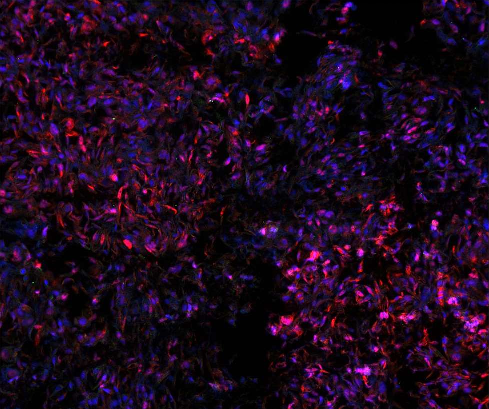
Immunofluorescence (IF) is a technique that permits visualization of virtually many components in any given tissue or cell type. This broad capability is achieved through the combination of specific antibodies labeled with fluorophores. Compared to immunohistochemistry (IHC), IF has superior sensitivity and signal amplification with various microscopy techniques.

Despite its wide range of applications, inexperienced researchers often face troubles in successfully detecting and analyzing clear fluorescent signals from their proteins of interest. The aim of our present paper is to provide a brief primer for researchers, especially those who just start using the IF method.
Sample preparation
Before starting an immunostaining, a literature study should be done to determine the expression level and intracellular localization of the protein of interest in the chosen model system.
IF protocols exist for a variety of different samples or specimens. The simplest and most commonly used method is staining of cultured cells, which is also known as immunocytochemistry (ICC). In IHC, on the other hand, the presence of a protein or molecule is examined in a tissue-specific context. In addition to the preparation of tissue sections, it is also possible to perform IF with whole organisms, a process referred to as "whole mount IHC".
Sample fixation
Fixation is the first step in an IF procedure and aims to maintain cells or tissues in their current state over an extended period. Different fixation methods are useful for IF, and each reagent affects the epitope of the primary antibody differently. Antibody binding sites can be masked or disrupted by fixation, which impairs the quality of IF staining. Since antibody binds differently to its antigen dependent on the various fixation compounds, it is necessary to try several different fixation methods to obtain better staining results.
Permeabilization
Permeabilization is required to allow penetration of probes into fixed cells or tissues. Triton X-100 and Tween-20 are non-ionic detergents commonly used for IF permeabilization. This step must be optimized based on the protein of interest, the used antibody, and the experimental conditions. Especially when staining membranous proteins, the permeabilization step has to be done with caution, as Triton X-100 can disrupt cell membrane.
Other permeabilization agents may be useful for specific applications. Digitonin has been used to selectively permeabilize the plasma membrane while leaving intracellular membrane intact. Saponin has been used for reversible.
Blocking
Protein solution or normal serum is used to dilute the antibody for binding as well as to block non-specific binding sites on cells prior to the antibody incubation step. Traditionally, normal serum from the same host as the secondary antibody is used for blocking in IF protocols. However, purified proteins like bovine serum albumin (BSA) or gelatin at 1-5% are also effective blocking agents.
Direct or indirect IF
There are two classes of IF techniques, direct and indirect.
Direct IF uses a single antibody chemically linked to a fluorophore. The antibody recognizes the target molecule and binds to it, and the fluorophore it carries can be detected via microscopy. Due to the direct conjugation of the antibody to the fluorophore, this technique has several advantages over the indirect protocol below. It reduces the number of steps in the staining procedure, making the process faster and can reduce background signal by avoiding some issues with antibody cross-reactivity or non-specificity. However, since the limited number of fluorescent molecules that can be bound to the primary antibody, direct IF is less sensitive than indirect IF.
Indirect IF uses two antibodies, an unlabeled primary antibody specifically binds the target molecule, and a secondary antibody, which carries the fluorophore, recognizes the primary antibody and binds to it. Multiple secondary antibodies can bind a single primary antibody. This provides signal amplification by increasing the number of fluorophore molecules per antigen. This protocol is more complex and time-consuming than the direct protocol above, but it allows for more flexibility since a variety of different secondary antibodies and detection techniques can be used for a given primary antibody.
Multiplex IF
Speaking of different colors, having a palette of secondary antibodies in several different species and colors makes it easy to perform multiplex IF. By using multiple primary antibodies from different species on a single sample, you can then use corresponding secondary antibodies conjugated to distinct fluorophores to visualize multiple targets at once. An advantage of multiplexing is that you can ask how multiple proteins relate to one another in the same sample. For this strategy to work, it is important to ensure that the excitation/emission spectra of the fluorophores you are using do not overlap. Multiplex IF requires some additional optimization as each antibody performs differently and some antibodies can impact the performance of others. But, once you do find the right protocol, multiplex IF can give you impactful and beautiful results.
Nucleus staining and sample mounting
The immunoreaction is often followed by a nucleus staining with DNA dyes. It enables better orientation within the cell or tissue sections during microscopy. Dyes such as Hoechst or DAPI, which enter the nucleus even without permeabilization and intercalate into the DNA, are used for this. For this reason, you should be very careful to avoid direct skin contact with these dyes!
Mounting medium is a viscous liquid that allows coverslips to be mounted on specimens on glass slides. This keeps the samples from drying out and allows imaging with oil immersion objectives. Since the excitation light used in fluorescence microscopy can cause fluorophores to photobleach, antifade compounds are often included in the mounting medium to prevent signal loss during imaging.