Traditional RNA detection methods like qPCR and RNA-seq offer quantitative information on gene expression, however they lose the spatial information from tissues, which eliminates critical details such as tissue heterogeneity and tissue microenvironment. Immunohistochemistry (IHC) can localize protein expression but is limited by antibody specificity, sensitivity, and challenges in detecting secretory proteins.
RNAscope, an advanced cellular and molecular diagnostic, is considered the gold standard in next-generation RNA in situ hybridization (ISH) due to its patented double-Z probe design and a cascading signal amplification system. This system gives it an unparalleled ability to balance single-molecule sensitivity with high specificity. As a result, the technology overcomes the limitations of traditional ISH and can detect multiple RNA targets in intact tissue sections. The platform is also compatible with both fluorescence and chromogenic imaging modalities, offering a revolutionary tool for spatial gene expression analysis.
Technical Advantages
- Single Molecule Resolution: Localizes and counts individual RNA molecules for each RNA target
- Ultra-High Spatial Resolution: Preserve tissue architecture, see the spatial distribution of gene expression
- Broad Sample Types: Formalin-fixed paraffin-embedded (FFPE) tissues, fresh frozen tissues, smears, cultured cells, etc.
- High Specificity and Low Background: Patented double-Z probe design greatly reduces non-specific hybridization
- Multiplex Target Detection: 2-plex, 4-plex, or even 12-plex targets can be detected simultaneously.
Detection Any RNA
Our RNAscope service covers the following research directions and target types:
- Targeting various RNA types, including mRNA, lncRNA, miRNA, and more.
- Common species such as human, mouse, rat, primate, and support for non-model species detection.
- Co-expression analysis of cell markers in the tumor microenvironment.
- Study of RNA expression and its spatial association with tissue architecture in disease.
Our RNAscope Service
Custom probe design
We can design probes based on known database IDs or based on client provided custom reference sequences.
Probe design strictly follows technical standards for both target specificity and signal intensity.
RNAscope detection service
Comprehensive experimental outsourcing service: sample preparation → hybridization → staining and imaging.
Flexible staining options (chromogenic or fluorescence multiplexing).
Optional additional services: tissue section preparation, tissue scanning, and quantitative image analysis.
Workflow
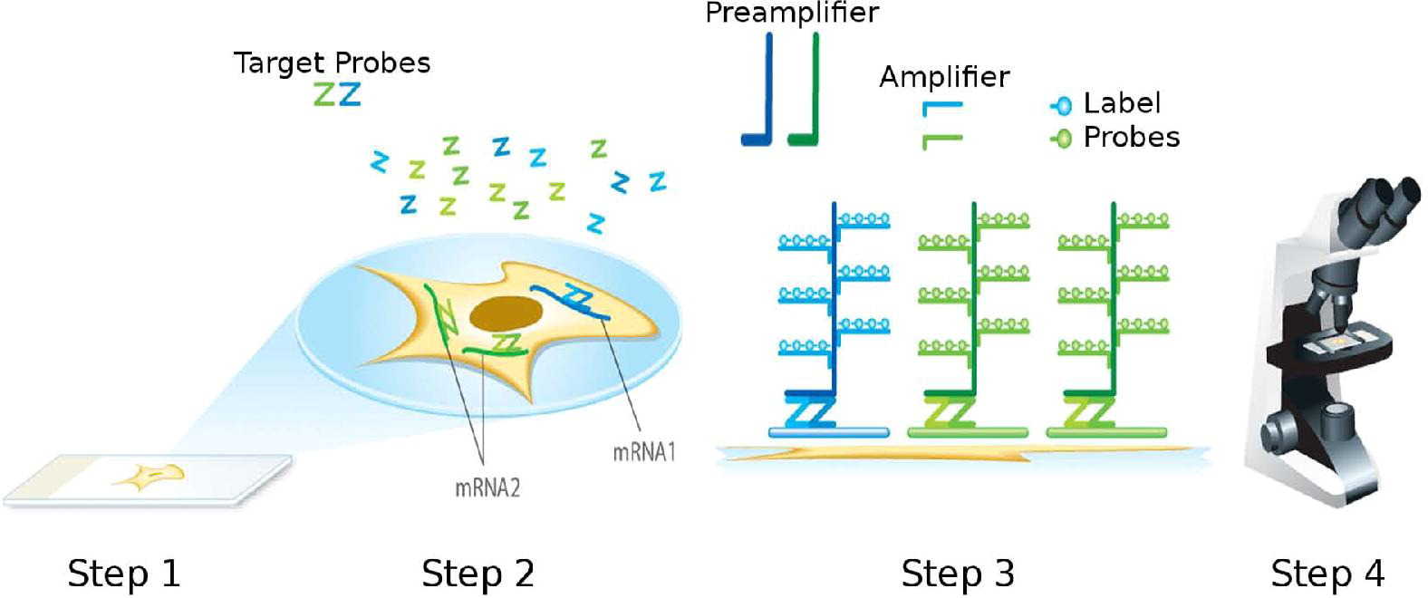 Fig. 1. Schematic of the RNAscope assay procedure (Wang F, Flanagan J, et al., 2012).
1. Sample Reception and Preprocessing: Receive tissue sections or cell samples from the client, perform quality control and necessary preprocessing (e.g. deparaffinization, rehydration, protease digestion) to ensure sample suitability for RNAscope detection.
2. Probe Hybridization: Adjust hybridization temperature and time to ensure full probe binding to target RNA when combining custom-designed probes with RNA samples.
3. Signal Amplification and Detection: Amplify the signal using RNAscope's patented amplification system and visualize target RNA signals using chromogenic or fluorescent labeling.
4. Microscopy and Image Acquisition: Observe the samples using high-resolution microscopy, capturing clear images that document the spatial distribution of gene expression.
5. Data Analysis and Reporting: Perform quantitative analysis of the acquired images, measure gene expression levels and distribution patterns, and provide a detailed experimental report.
Core Features
- One-Stop Service: Complete service from probe design, experimental operation, imaging, and data analysis.
- High-Quality Standards: Adherence to RNAscope's standard operating procedures (SOP) throughout the process.
- Diverse Sample Experience: Supports a wide range of sample types, from FFPE and frozen tissues to cell smears.
- Experienced Technical Support: Expert molecular biology and pathology team with experience in custom service and problem solving.
Featured Applications
- Spatial Biology: Characterize the spatial expression pattern of genes of interest in tissues.
- Biomarker Validation: Tissue localization and level-based validation of RNA biomarkers.
- Gene and Cell Therapy: Verify expression of therapeutic genes in target tissues.
- Stem Cell Research: Monitor the dynamic expression of genes of interest during stem cell differentiation.
- Neurobiology: Localize gene expression in the nervous system, and analyze the differences of functions among regions.
- Infectious Disease Research: Spatial detection of pathogens RNA and host response patterns.
Reference
- Wang F, Flanagan J, et al., RNAscope: a novel in situ RNA analysis platform for formalin-fixed, paraffin-embedded tissues. J Mol Diagn. 2012. 14(1):22-9.