Why Fixation?
Fixation is the first key factor to ensure the scientific value and quality of biological specimens. It is important for at least 3 purposes:
1) To stop tissue autolysis by hydrolytic enzymes released from cells, and thus to better preserve cellular morphology for analysis.
2) To immobilize tissue and cellular antigens for immunolabeling of these antigens, and
3) To offer better preparation of samples for histological sections.
It is therefore recommended that tissue be fixed as soon as possible.
The Types of Fixation
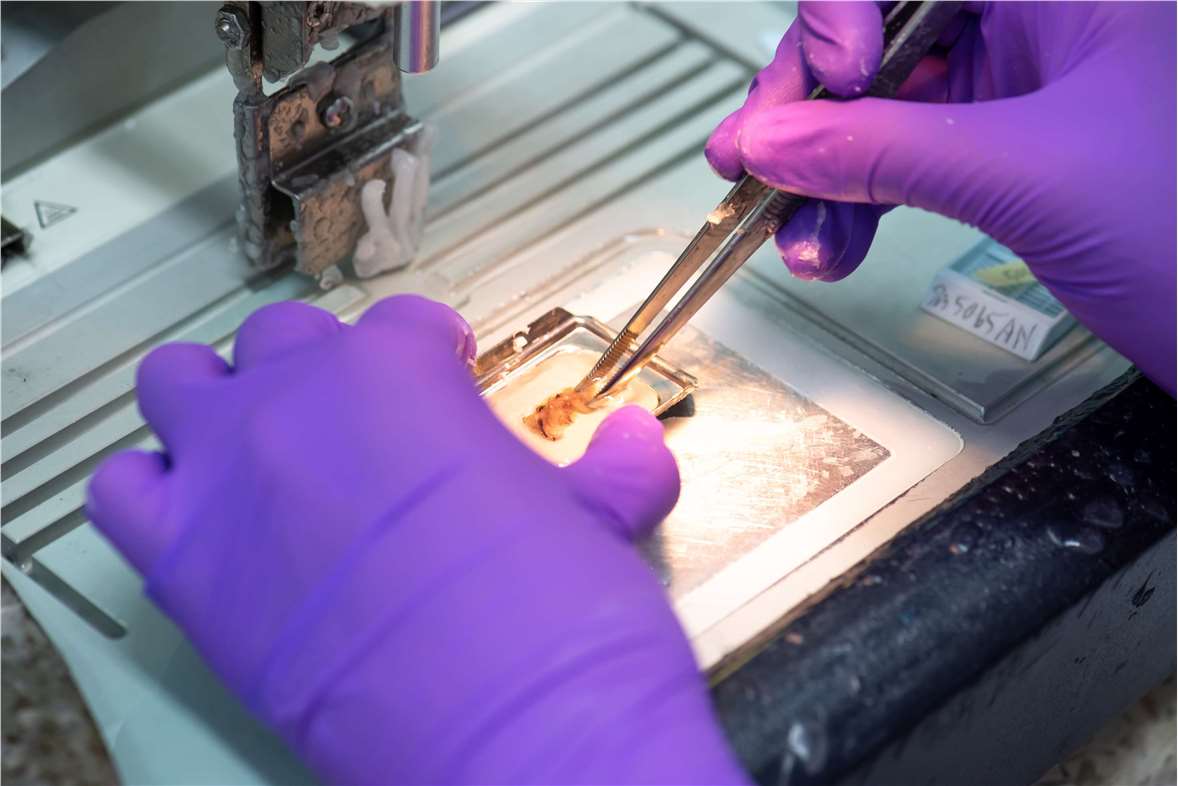
Fixation of tissues can be achieved by physical or chemical means.
Physical methods include heating, microwaving and cryopreservation. Heat fixation is rarely used on tissue specimens, its application being confined to microorganism smears. Microwave fixation, which can be considered as a form of heat fixation, is now widely practiced in routine laboratories. Cryopreservation, usually in the form of freeze drying, has some applications in histochemistry but is not usually applied to diagnostic tissue specimens.
Chemical fixation is usually achieved by immersing the specimen in a fixative (immersion fixation) or by perfusing the vascular system with a fixative (perfusion fixation). For some specialized histochemical procedures, fixatives are occasionally applied in vapor form.
The Mechanism of Fixation
The two main mechanisms of chemical fixation are cross-linking and denaturation.
Cross-linking: Non-coagulant fixing agents chemically react with proteins and other cell and tissue components, becoming bound to them by addition and forming intermolecular and intramolecular cross-links. This can have an effect on the subsequent staining of a particular protein as well as altering its molecular conformation and thus its solubility.
Denaturation: This effect is induced by dehydrants such as the alcohols or acetone. These reagents remove and replace free water in cells and tissues and cause a change in the tertiary structure of proteins by destabilizing hydrophobic bonding.
Factors Involved in Tissue Fixation
Time interval: The time interval from removal of the tissue to fixation is also important. The faster you can get the tissue and fix it, the better.
Specimen dimension: Penetration of tissues depends on the diffuse-ability of each individual fixative, which is usually slow. One way to address this problem is to section the tissues thinly (2 to 3 mm).
Concentration of fixative: The concentration of fixative should be adjusted down to the lowest level possible, too high a concentration may adversely affect the tissue and produce artefact similar to excessive heat.
Volume ratio: The volume of fixative is important. There should be a 10:1 ratio of fixative to tissue. One way to partially solve the problem is to change the fixative at intervals to avoid exhaustion of the fixative. Agitation of the specimen in the fixative will also enhance fixation.
Buffering: Fixation is best performed near neutral pH, in the range of 6-8. Hypoxia of tissues lowers the pH, so buffering capacity must be present in the fixative to prevent excessive acidity.
Temperature: In general, an increase in temperature increased the rate of fixation but also increased the rate of autolysis and diffusion of cellular elements. Traditionally, 0 to 4°C has been considered the ideal temperature or the fixation of specimens. Now fixation is routinely carried out at room temperature.
pH: Good fixation is achieved at a pH of 6-8. Outside this pH there may be damage to the ultrastructure.
Duration: Primary fixation in buffered formalin for 2-8 hours (<24 hours). Prolonged fixation causes hardening and shrinkage of tissue.
The Classification of Fixatives
There are five major groups of fixatives, classified according to mechanism of action:
1) Aldehydes (Formaldehyde/10% Formalin/Glutaraldehyde)
Tissue is fixed by cross-linking formed in the proteins, particularly between lysine residues. This cross-linking does not cause much damage to the structure of the protein, so antigenicity is not lost.
Formaldehyde is friendly to immunohistochemistry. Formalin penetrates tissue well, but is relatively slow. Glutaraldehyde causes deformation of alpha-helix structure in protein, which is not conducive to immunohistochemical staining. However, it fixes very quickly so is good for electron microscopy. It is poorly penetrating but gives the best overall detail of the cytoplasm and nucleus.
2) Mercurials (Mercuric chloride/B-5/Zenker's)
Mercurials fix tissue by an unknown mechanism. These fixatives penetrate relatively poorly and cause some tissue hardness, but are fast and provide excellent nuclear detail. Their best applications are for fixation of hematopoietic and reticuloendothelial tissues.
The mercuric-containing fixatives, such as B-5 and Zenker's, are rarely used in current practice and are thought to function by binding to sulphydryl and amino groups in additive reactions.
3) Alcohols (Carnoy's/Methanol/Ethanol)
Alcohols, which are protein denaturants, are generally not used for tissues because they cause too much brittleness and hardness. However, they are very good for cytologic smears as they act quickly and give good nuclear detail.
Carnoy's fluid is generally used for specific purposes. Ethyl alcohol is used as a fixative for enzymes.
4) Oxidizing agents (Permanganate fixatives/Dichromate fixatives/Osmium tetroxide)
Oxidizing agents cross-link proteins, but cause extensive denaturation. Some of them have specialized applications, but are used very infrequently.
5) Picrates (Bouin's)
Picrates include fixatives with picric acid. They react with histones and basic proteins to form crystalline picrates with amino acids. This is a very good preservative for glycogen.