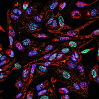
Immunofluorescence (IF) is a powerful tool for detecting and identifying the subcellular distribution and movement of proteins and other molecules within cells and tissues. IF can be used as an alternative to traditional chromogenic labeling used in Immunohistochemistry (IHC) or Immunocytochemistry (ICC). By using multiple secondary antibodies, the colocalization of proteins of interest can be studied, which cannot be achieved through conventional IHC or ICC. This is an advantage when analyzing many targets at the same time, generating a high volume of accurate data.

While IF is a well-established and widely adopted technique, common problems such as background interference, lack of specificity, weak signal, or poor reproducibility may encounter, when working with biological specimens. Here we present some helpful tips and tricks for improving IF staining results.
Non-Specific Staining
| Problem | Solution |
| Spectral overlap | If imaging more than one fluorescent probe, the fluorophores may have excitation and emission spectra that overlap. Adjust your light sources and filters to pick up only one signal at a time. If this is not possible, choose new fluorophores that do not have spectral overlap. |
| Primary antibody raised again the same species as tissue being analyzed (e.g., mouse on mouse) | Try using a primary that is raised against a different species. Otherwise try to block the endogenous IgG with serum from the same species as the secondary. Incubate sections with 1% Triton to clean the tissues. Or Use TBS-Tween 20 as a washing buffer, rather than using PBS-Tween 20. |
| Aggregates | Spin down secondary antibodies in a microcentrifuge to move aggregates to the bottom of the tube. |
Weak or No Staining
| Problem | Solution |
| Very low or no antigen expression. | Run a positive control. If the protein is present, but not abundantly, use an amplification step to maximize the signal. |
| Damaged epitope. | Reduce fixation time or change to another fixative. |
| Wrong permeabilization method. | Optimize or skip the permeabilization step. |
| Tissue/cells dried out. | Always keep the sample moist. |
| Fluorescent tag bleached. | Perform incubations and store samples in the dark. Samples may be mounted in an anti-fade solution. Samples should be imaged immediately following mounting for the best results. |
| Inappropriate primary antibody. | Confirm that the antibody has been validated in IHC, and specifically what type - formalin fixed, paraffin-embedded, fresh frozen, etc. Test the antibody in a western blot to make sure it hasn't been damaged. |
| Primary and secondary antibodies incompatible. | Choose a secondary antibody raised against the host species of the primary antibody. For example, if the primary antibody was raised in mouse, select an anti-mouse secondary antibody. |
| Too high primary/secondary antibody dilution, too short incubation time, too low incubation temperature. | Increase the antibody concentration or the incubation time/temperature |
| Inappropriate microscopy detection method. | Use a more sensitive method for image acquisition. Check your used filter/laser setup. Use a brighter fluorophore for detection. |
High Background
| Problem | Solution |
| Inappropriate or too long fixation | Reduce fixation time or change the fixative. |
| Insufficient blocking | Increase the blocking incubation period and consider changing the blocking agent. Usually, a blocking serum matching the species of the secondary antibody is recommended. |
| Not enough washing | Verify that all washing steps are carried out properly. If necessary, repeat or prolong the washing step. |
| Tissue too thick | Consider using thinner tissue sections. |
| Samples dried out. | Always keep slide moist. |
| Too high primary/secondary antibody concentration, too long incubation time, too high incubation temperature. | Optimize the antibody concentration and incubation time/temperature, consult manufacturer's protocol. |
| Non-specific binding of secondary antibodies | Run a secondary control without the primary. If there is staining, then change the secondary. |
| Autofluorescence | Check the sample autofluorescence by using unstained controls. Avoid glutaraldehyde fixative or wash with 0.1% sodium borohydride in PBS to remove free aldehyde groups. May be due to endogenous molecules (FAD, FMN, NADH, lipofuscin), and will require pre-photobleaching, treatment with sudan black, or cupric sulfate. |