Immunohistochemistry (IHC) and immunocytochemistry (ICC) both utilize antibodies to provide visual details of protein abundance, distribution, and localization, but there are differences between them. As the name suggests, IHC is used to detect proteins in fixed tissues. ICC, on the other hand, is used to detect proteins in isolated cells. Both IHC and ICC are important in the fields of biology, biochemistry, genetics, microbiology, and medicine.
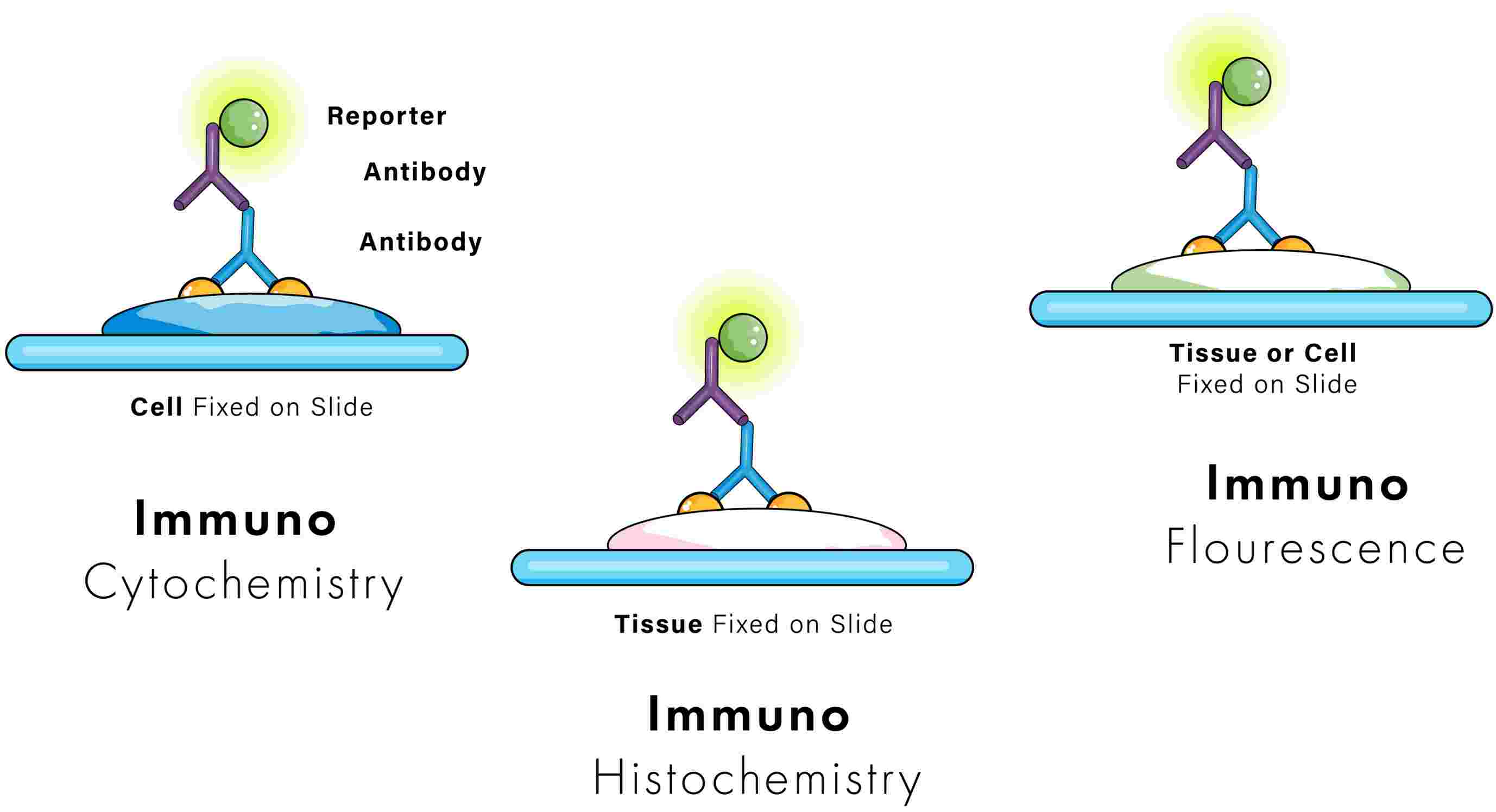
Sample Source
Prior to IHC, tissues are removed from patients or animals, either frozen or chemically preserved (fixed), and then embedded in paraffin wax. Sections as thin as 4 μm are sliced from frozen or paraffin-embedded tissue and mounted onto glass slides in preparation for antibody-mediated staining. In this way, researchers can observe the localization of target antigens within cellular components while maintaining the original architecture of the surrounding tissue.
The sample source for ICC can be any cell suspension obtained from a patient or animal (e.g., blood smears, swabs and aspirates) or cells cultured in monolayers, usually on sterile glass coverslips. The latter is the most commonly used sample type for ICC. ICC is also synonymous with IF staining, especially when we present the results with cultured cells. The two abbreviations are often used interchangeably in this context.
Both IHC and ICC begin with a fixation step, using formaldehyde or a similar compound to lock the sample in place. After fixation, the procedures diverge.
Sample Processing
Aside from biological sources, IHC and ICC differ in the amount of sample processing required before antibody-mediated staining. For example, ICC is associated with whole cells, which must be permeabilized by a fixation procedure or a separate permeabilization step to facilitate antibody penetration to the intracellular targets. Depending on the thickness of the section and the method of fixation, IHC samples may not have to undergo a separate permeabilization step. However, most formalin-fixed, paraffin-embedded (FFPE) IHC sections must be further processed prior to antibody staining to uncover latent epitopes on antigenic targets. This is usually referred to as epitope or antigen retrieval.
Labeling Method
IHC and ICC have traditionally used chromogenic reagents to detect target antigens. In chromogenic assays, enzymes like horseradish peroxidase (HRP) convert soluble substrates including 3,3'-Diaminobenzidine (DAB) and 3-amino-9-ethylcarbazole (AEC) into insoluble colored products at the antigenic site. In IF assays, the fluorochrome reagents label, either directly or indirectly, the primary antibodies and, once excited, will emit light at a specific wavelength.
With the extensive list of available fluorescent labels and their ease of use in multiplex applications, researchers are more often choosing ICC/IF and fluorescent IHC. Additionally, the use of fluorescent conjugated antibodies specific against organelle markers, facilitates the analysis of proteins of interest and extends the multiplex capabilities in both IHC and ICC.