What is FISH?
FISH, fluorescence in situ hybridization, is a cytogenetic technique enabling "mapping" of the genetic material of cells. It is commonly used to label DNA, providing information about the location, length, and copy number of a specific gene or chromosome portion. In addition, it can be applied to all types of RNA and is primarily used to detect the copy number and location of mRNA to visualize cellular transcription activity.
How does FISH Work?
1) Preparation of fluorescent probes
- A fluorescently labelled FISH probe is designed that is complementary to the DNA of interest.
- Size range from 20-40 base pair to 1000 bp.
- They are tagged with fluorescent dyes like biotin, fluracine and diaoxigenin.
2) Denaturation of probe and the target sequence
- The FISH probes and the slide are heat treated together to denature both the FISH probe DNA and the genomic DNA into single strands.
3) Hybridization of probe and the target sequence
- Formation of duplex between two complementary nucleotide sequence
- It can be between DNA-DNA, DNA-RNA and RNA-RNA.
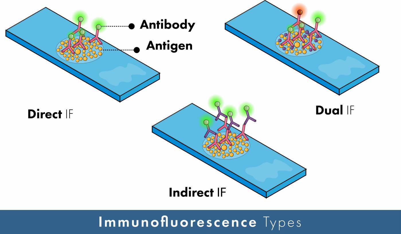
4) Detection
- Detection has Two types:
a) direct labelling - label is bound to the probe less sensitive.
b) indirect labelling - require an additional step before detection.
5) Visualization
- The slide is visualized under a fluorescent microscope and the labelled probe fluoresces.
- Multiple probes can be simultaneously hybridized and visualized by using probes with differently colored fluorescent labels.
Probes
Three different types of FISH probes are used. Each of them has a different purpose.
1) Whole chromosome probes - these probes are libraries of small DNA sequences which are complimentary for different sequences of a chromosome. These probes are used for the process of chromosome painting in order to identify the numerical aberrations in chromosomes.
2) Alphoid or centromeric repeat probes - they are complementary to the centromere region of the chromosome more specifically to the repetitive alpha satellite DNA sequences. Use of these probes let us know the number of chromosomes and thus the number of a specific chromosome.
3) Locus-specific probes - as the name suggest this type of probes are complementary to a specific position in the chromosome. These probes are useful in determining whether a gene has a normal copy number or not.
A number of things are considered during making the choice of a probe such as - sensitivity, specificity, the stability of hybrids, and ease of application.
The Advantages of FISH
1) It aids interpretation of results from karyotyping and arrays.
2) Metaphase FISH can provide positional information (this needs cell culture).
3) Interphase FISH can be performed on FFPE slides (it provides no positional information, but it can provide information on fusions, amplification and deletions without the need for cell culture).
4) It detects whole genome ploidy changes.
5) It detects mosaicism.
6) Probes are available for almost any genomic region.
The Limitations of FISH
1) Experienced analysts are needed to interpret FISH images.
2) It cannot detect uniparental disomy.
3) Atypical rearrangements may be missed, for example some t (15;17) fusions in hemato-oncology.
4) Co-localization can occur (signals from two FISH probes overlap because the chromosomes happen to be in the same place on the slide and appear as one).
5) Cross-hybridization can occur (the probe binds to regions with repetitive sequences).
What Information can FISH Actually Provides?
In medicine, FISH finds its best application in correlations between DNA sequences (mutations and modifications within then) and phenotypes (usually cancer or disease).
1) Simultaneous detection of aberrations on many different chromosomes: SKY is an improvement of the FISH application, based on the use of fluorescent probes of different colors in order to visualize and distinguish many different chromosomes. This is very important in the field of Gene Mapping.
2) Another improvement in the application of Gene Mapping is the ability to identify several different regions on the same chromosome by using different probes together. The process, known as Multiplex FISH, allows painting of the entire chromosome complement in a single hybridization by labelling each chromosome with a different combination of fluorophores.
3) Comparative genomic hybridization analyzes chromosomal imbalance in tumors and examines possible correlations between findings and tumor phenotypes.
4) The study of chromosomal aberrations also in non-dividing cells, leading to chromosomal mapping of some deleted or amplified region.
5) Visualization and measurement of telomere length.
6) Quantification of gene copy number and the amount of protein.