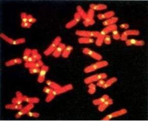
Fluorescence in situ hybridization (FISH) technology allows the detection of specific nucleic acid sequences in morphologically preserved chromosomes, cells, and tissues. The unambiguous detection of structural or copy number changes of whole chromosomes or chromosome specific regions is an important predictive and prognostic factor in human disease. A wide variety of sample types are amendable to FISH, making it particularly valuable in the study of archival materials, such as formalin-fixed paraffin-embedded tissues.
Creative Bioarray offers FISH services for a variety of specimen types, including non-paraffin, paraffin and paraffin tissue microarrays. Our FISH services can be performed to enumerate the copy number of cancer-related genes and patterns of gene-related gains and losses; map the location of DNA sequences, genes or transgene insertions; and determine the presence or absence of interspecies cells in xenograft or chimeric animal models. We are happy to share simple but useful tips to improve your daily tasks as well as the overall quality of your results. With this in mind, below is a list of tips for achieving high-quality data by FISH.
Sample Preparation Tips
- Positively charged slides should only be used for FFPE sections. If these slides are used for cell suspensions, they may produce a high green background.
- When using an enzyme solution to digest FFPE slides, be sure to use the enzyme liberally as evaporation may cause the enzyme to recede from the edges, resulting in inconsistent digestion.
- After pre-treatment, consider using DAPI to assess for over/under digestion of FFPE sections, this can be washed off before applying the FISH probe.
- Consider using a mild hypotonic treatment before fixing CD138+ selected cells, which may improve cell morphology.
- If your cells appear intact with a three-dimensional look to them, and there is visible cytoplasm using a phase contrast microscope, you can try re-fixing the cells in 1:1 methanol:acetic acid.
- Consider using Pepsin to digest excess cytoplasm and cell debris in Cytospin samples to improve hybridization quality.
- Try pre-fixing bone marrow or peripheral blood samples by slowly adding ice-cold fixative immediately after the hypotonic treatment to improve cell preparation.
- When using archived cell pellets, consider re-fixing using fresh fixative to ensure optimal results.
- Try to avoid baking or aging of slides (if not required for other applications such as g-banding) as it may reduce signal fluorescence.
- Try cleaning the slides with 70% ethanol before use, to get rid of any dust or debris. A high degree of debris may lead to a high background.
- Perform tissue enzyme digestion on a hotplate at 37°C for best results.
Probe Application Tips
- When applying FISH probes, try not to push down too hard on the coverslip as it can squeeze out of the sides, causing a patchy hybridization.
- To avoid patchy hybridizations, be sure to press out visible bubbles when applying the coverslip on the probe mixture.
- Make sure the pipette tip is long enough so that the pipette itself doesn't come into contact when the tip reaches the bottom of the vial. Pipettes entering a probe vial may carry over probe reagent from one vial to the next resulting in cross contamination.
- If your chromosomes appear distorted, it may be because the coverslip is not sealed. To improve chromosome morphology, try sealing the coverslips prior to hybridization.
- Filtered pipette tips can reduce the intermittent background problem from debris being expelled onto FlSH slides.
- When making FISH slides from fixed cell suspensions, use a template to ensure that the suspension is consistently spotted in the same place. Use the template for probe application also, which ensures the FlSH probe is applied in the correct place.
Denaturation Tips
- Use calibrated temperature - measuring devices to regularly QC hybridization units, there may be significant slot to slot variability that can get worse over time, leading to poor results.
Hybridization Tips
- Test ramp up and ramp down temperature times periodically to ensure they are within manufacturer specifications.
- Maintaining a humidity strip on automated hybridization units according to the manufacturer's recommendations helps ensure optimal results from these units.
- Automated hybridization units may fail to provide sufficient humidity, resulting in poor hybridization. Try using a humidified chamber to attain consistently higher humidity levels.
- If you are using an incubator for hybridization, use a darkened, humidified chamber which has been pre-moistened (wet tissue) and pre-warmed.
Post Hybridization Wash Tips
- Always balance the pH of wash solutions during preparation, and periodically during use to ensure they produce optimal results.
- Try rinsing slides in a dehydration series of EtOH washes (70%, 85%, and 100%) after the room temperature washes to reduce background.
- When measuring the temperature of wash solutions, be sure to do this inside the jar to replicate the correct conditions.
Probe Analysis Tips
- Many types of fluorescence microscope filter degrade over time and should be treated as consumables. Ensure regular maintenance and replace when necessary.
- Liquid light-guides in modern fluorescent light sources will degrade over time, resulting in reduced light transmission, and should be treated as consumables. Make sure there are no sharp bends in the light-guide, schedule regular maintenance and replace when necessary.
- Try to use just one type of immersion oil in your laboratory, different types of oil may be immiscible.
Reagent Storage Tips
- When not in use, ensure that the enzyme solution is correctly stored at 2-8℃ to preserve the enzyme activity.
- Keep light exposure of the probe to a minimum as this may lead to photodegradation.
- Aliquot probes into smaller vials to avoid excessive light exposure. Only take the probe to be used from the freezer instead of all of the probes to minimize light exposure/freeze thaws.